Introduction
Physiotherapy in Toronto for Vertigo
Welcome to In Balance Physiotherapy's resource on Vertigo.
We have all experienced, for a variety of reasons, the sensation of being dizzy. Dizziness is a catch-all term that can describe an assortment of symptoms from feeling woozy after a ride at the amusement park or feeling light-headed as you stand up from sitting, to a sensation of being unstable or feeling like you are going to faint. Dizziness as a medical complaint is very common. Some people, however, suffer with a more severe and specific type of dizziness called vertigo. Vertigo is the medical term used to describe a patient’s symptom of perceiving (falsely) that they are moving within their environment or that objects in their environment are moving around them. This movement is generally described as a whirling, spinning or rotating type of motion.
This guide will help you understand:
- what tests your healthcare professional will do to diagnose it
- what In Balance Physiotherapy’s approach to rehabilitation is
Anatomy
The anatomy and physiology underlying the human body’s sensation of balance is complex. Many systems are involved including the brain, the spinal cord, the eyes, the ears and the receptors in the skin, joints and muscles. Disruption to any of these areas through injury or disease can affect one’s feeling of being balanced. Vertigo is the chief complaint of patients when there is dysfunction or disease within the inner ear portion of the balance pathway or along the inner ears’ connections to the brain.
The inner ear, which is also called the labyrinth of the ear, is made up of three primary structures which moderate balance and equilibrium; the semicircular canals along with the saccule and utricle. Collectively this system of the inner ear is termed the vestibular system or vestibular apparatus. The inner ear also contains the cochlea, which is the main structure involved in hearing.
The three semicircular canals work to detect rotational motion of the head. The canals are positioned at 90-degree angles to one another and are filled with fluid called endolymph. Hair cells are located at the base of each canal and project up into the endolymph. Movement of the head causes movement of the endolymph within the canals, which in turn causes the hair follicles to move accordingly and emit impulses about the position of the head in space. Hair follicles in the saccule and utricle add to the balance information by providing feedback about the position of the head in reference to gravity (vertical orientation) as well as detecting linear motion of the head.
Sensory information from the inner ear is relayed to the brain via the vestibular portion of the eighth cranial nerve (CNVIII), which is also called the vestibulocochlear nerve. The cochlear portion of the nerve transmits information about hearing. Specific areas of the brain, in particular the cerebellum and brain stem as well as portions of the cortex, process the inner ear sensory information. When both the right and left inner ears are sending the same information, the brain processes that the body is balanced. When the body or head moves, the sensory input from the ears is not identical so the brain perceives motion and the body adjusts accordingly.
The ears work in close relation with the eyes in order to maintain equilibrium and balance. The vestibulo-ocular reflex is an automatic function of the eyes, which stabilizes images on the retina in response to the vestibular sensory input from the ears. This reflex causes the eyes to move in the opposite direction to the movement of the head in order for the eyes to remain fixed on a target. Thus, accurate vestibular input from the ears affects how the eyes adjust, and to one’s sense of being balanced. The accurate relay of information from the eyes along the cranial nerve called the optic nerve (CN II) to the brain is also required.
For the healthcare professional, assessing reflexive eye motion is important in order to determine whether the vestibular system is working properly. If one inner ear is affected by disease or injury then the sensory input being sent to the brain will falsely indicate movement from that vestibular system. In this case the eyes will adjust accordingly and move opposite to the perceived motion despite the head actually being still. An involuntary back and forth movement of the eyes results. This movement of the eyes is called nystagmus. Nystagmus can be caused by several reasons other than vestibular problems, however in the case of accompanying vertigo, nystagmus leads the health care professional to the suspicion that the vestibular system is the culprit.
The brain amalgamates the vestibular information from the inner ears with sensory information from the eyes as well as the information coming from the receptors in the muscles and joints to provide the body with its overall sense of balance within its environment.
Causes
Disruption along any portion of the anatomical pathway described above can affect one’s perception of balance or equilibrium. A problem with the inner ear portion of the pathway or the sensory information being relayed to the brain via the vestibulocochlear nerve is termed a peripheral vestibular disorder. If the problem affecting one’s balance is due to damage of a structure within the brain itself, which then affects the reception and integration of balance information, it is termed a central vestibular disorder.
Peripheral or central vestibular disorders can both cause vertigo. Some cases of vertigo may be due to both peripheral and central vestibular disorders.
The most common peripheral vestibular disorders causing vertigo are benign paroxysmal positional vertigo, Meniere’s disease, and vestibular neuronitis/neuritis or labyrinthitis. Other causes that will be discussed in this guide are migraine associated vertigo, acoustic neuroma, and vertigo as a symptom of Multiple Sclerosis.
Benign Paroxysmal Positional Vertigo
Benign paroxysmal positional vertigo (BPPV) is a common clinical disorder of balance, which is characterized by recurrent vertigo spells that are brief in nature (usually 10-60 seconds) and are most often triggered by certain head positions. Benign, in medical terms, means it is not threatening to life. Paroxysmal means it comes with a rapid and sudden onset or increase in symptoms.
BPPV is the most common cause of recurrent vertigo. The cause of BPPV is proposed to be calcium carbonate crystals (called otoconia or otoliths), which are sometimes termed ‘ear rocks’ within the semicircular canals of the inner ear. Usually these crystals are located within the utricle and saccule of the ear. It is thought that these crystals dislodge and migrate to the semicircular canals of the ear. The cause of this dislodgement is postulated to be a number of possible reasons such as an ear or head injury, an ear infection or surgery, or from natural degeneration of the inner ear structures. Often a direct cause cannot be identified.
The otoconia settle in one spot in the canal when the head is still. The most common canal for settlement in is the posterior semicircular canal. A sudden change in head position, often brought on by activities such as rolling over in bed, getting out of bed, bending over, or looking upwards, causes the crystals to shift. This shift in turn sends false signals to the brain about equilibrium, and triggers the vertigo.
Vertigo due to BPPV can be severe and accompanied by nausea. The attacks can occur seemingly for no reason and then disappear for weeks or months before returning again. Generally BPPV affects only one ear and although it can occur at any age it is often seen in patients over the age of 60 and more often in women. Nystagmus is usually present.
Vestibular Neuronitis or Labyrinthitis
Vestibular neuronitis or labyrinthitis is an inflammation of the inner ear or its associated nerve (the vestibular portion of the vestibulocochlear nerve), which causes vertigo. Hearing may also be affected if the infection affects both portions of the vestibulocochlear nerve.
The vertigo caused by vestibular neuronitis or labyrinthitis is of a sudden onset and can be mild or extremely severe. Nausea, vomiting, unsteadiness, decreased concentration, nystagmus and impaired vision may also accompany the vertigo. Most often the infections that cause inflammation of the inner ear or the vestibulocochlear nerve are viral in nature as opposed to bacterial. Proper diagnosis of the cause is important in order to provide the most effective and appropriate treatment.
Meniere's Disease
Meniere's disease is a chronic incurable vestibular disorder characterized by symptoms of episodic severe vertigo, fluctuating hearing loss, ear ‘fullness’ and/or ringing in the ear (tinnitus), and nystagmus.
This disease derives its name from a French physician, Prosper Meniere, who theorized in the late 1800’s about the cause of this repertoire of symptoms, which he noted in many of his patients.
Early-stage acute attacks of Meniere’s disease vary in their length anywhere from 20 minutes to 24 hours. The attacks can occur regularly within a week or may be separated by weeks or months. Other symptoms may coincide with the attack such as anxiety, diarrhea, trembling, blurry vision, nausea and vomiting, cold sweats, and a rapid pulse or heart palpitations. Following the attacks patients often feel extreme tiredness, which requires many hours of rest to recover. For some patients time between attacks may be symptom free but other patients report ongoing related symptoms even between attacks.
The exact cause of Meniere’s disease is still not certain but it is theorized that it is due to an abnormal amount of endolymph fluid collecting in the inner ear and/or an abnormal buildup of potassium in the inner ear.
Migraine Associated Vertigo
Some patients who suffer from migraines (approximately 25-35%) experience migraine associated vertigo (MAV). MAV, (also called a vestibular migraine), may also be accompanied by nausea or vomiting and may last a few seconds or a few days. Other vestibular symptoms may also be noted in association such as motion intolerance, sensitivity to head movement, dizziness, a feeling of pressure in the ears, imbalance and spatial disorientation. With MAV the symptom of vertigo may precede the onset of the migraine or may appear as the headache pain develops. Vertigo may also occur during a headache-free time frame. Some patients will also present with a true BPPV after the migraine event has ceased.
Acoustic Neuroma
An acoustic neuroma is a benign (non-cancerous) tumour on the vestibulocochlear nerve. Early symptoms are related to loss of hearing in the affected ear, ringing in the ear (tinnitus), dizziness, and a feeling of fullness in the ear. The tumour is slow growing so symptoms come on gradually and may be easily overlooked in the early stages. As the tumour grows it may push on other nerves in the area and symptoms such as headaches or pain and numbness in the face may appear. Vertigo or other balance issues may arise with growth of the tumour.
Vertigo as a symptom of Multiple Sclerosis
Multiple Sclerosis (MS), which causes a demylenation of nerves, primarily attacks the cerebellum of the brain, as well as the brain stem (including the cranial nerves such as the vestibulocochlear nerve), and the spinal cord. The cerebellum is particularly important in regards to balance as it helps to integrate information received by the brain in order to both maintain balance and arrange coordinated movements. Damage to either the cerebellum and/or the vestibulocochlear nerve due to MS can cause vertigo.
Approximately 20% of MS sufferers will experience vertigo as a symptom. The vertigo attacks associated with MS can be short-lived or last for days or weeks at a time. A much more common symptom of MS sufferers rather than vertigo is general dizziness or lightheadedness.
Other causes of Vertigo
Although most cases of vertigo are related to peripheral or central vestibular disorders, other causes of vertigo may be identified such as alcohol intoxication, metabolic disorders, bacterial or viral infections, side-effects from medications, or side effects from overexposure to specific chemicals (ototoxicity). Even severe emotional issues causing anxiety can manifest in vertigo. In some rare cases, however, a cause for the symptom of vertigo goes unknown.
Related Symptoms
Vertigo is rarely experienced independent of other symptoms. Depending on the cause of the vertigo the following symptoms may accompany a vertigo attack or be experienced in close proximity, either before or after the attack, or as a result of the vertigo attack itself:
- dizziness
- headaches
- nausea and/or vomiting
- cold sweats
- ringing in the ears (tinnitus)
- fatigue
- hearing loss
- twitching eyes
- nystagmus
- ear pressure
- panic attacks
- a feeling of being pulled to or leaning to one side
- fear of falling
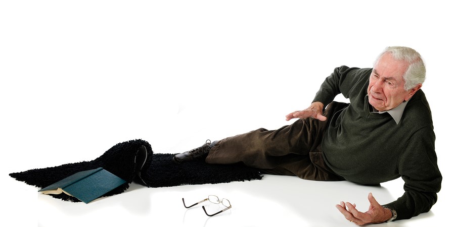
- increased risk of falls
- anxiety
- depression
- work difficulties
- decreased engagement in social activities
- decreased motion of the neck or body or avoidance of certain movements in order to avoid onset of an attack or limit symptoms
Diagnosis
A detailed history of your problem is the most crucial information needed by your healthcare professional in order to diagnosis true vertigo from general dizziness, to determine a cause for the vertigo, and then to implement appropriate treatment.
Your healthcare professional will ask you to describe your symptoms in detail. As explained above, true vertigo is more than just a general feeling of dizziness or lightheadedness, but rather a false sensation of spinning or rotating within your environment or your environment spinning or rotating around you.
Your healthcare professional will want to know when the first episode of your vertigo occurred, how long it lasted, and if it was associated with any other events such as a car accident, head trauma, or an illness or infection.
They will also want to know how often you have experienced the vertigo since the first episode, and the general pattern of symptom frequency. Your healthcare professional will ask if anything in particular triggers your symptoms such as moving your head in a certain direction or getting out of bed. They will also inquire about any other related symptoms such as nausea, vomiting, falls, visual disturbances, feelings of fullness or pressure in the ear, ringing in the ear, hearing loss, headaches or if you have a history of migraines. Your healthcare professional will also want to know if there is anything that makes your symptoms better, if you are taking any medications, or if you have a family history of any inner ear disorders or central nervous disorders.
Your healthcare professional will then perform a physical examination. They will look in your eyes to note any nystagmus and may do a basic examination of your ears by looking into them. A general examination of your balance will be completed and they may ask you to lie down and then get up from the lying position in order to determine if it brings on your symptoms. A general examination of the joints of your neck will also be completed to rule out any symptoms coming from the neck region. Other general physical examinations such as blood pressure in both lying and standing may also be assessed.
Dix-Hallpike's manoeuvre
Dix-Hallpike’s manoeuvre is a test that can be done by your healthcare professional in their clinical setting. This tests helps to determine if certain head movements are the cause of your vertigo. If positive this test can also determine which ear is the problem.
During this test your healthcare professional turns your head to one direction then assists you to quickly lie back while maintaining your head position and also hanging your head over the edge of the bed. Your healthcare professional watches your eyes for whether nystagmus occurs and also assesses the direction and quality of it.
After you sit upright for a few minutes to allow recovery, the same test is done with the head turned to the opposite direction.
The tests that can be done in your healthcare professional’s clinic in order to definitively determine the cause of your vertigo are limited. Depending on what your health care professional finds on their initial examination they may send you for a battery of other tests to further determine the cause of your vertigo.
Electronystagmography
Nystagmus, as previously explained, is an involuntary movement of the eyes. Nystagmus can indicate a problem with your balance system, particularly the nerve that runs from your ear to your brain (vestibulocochlear nerve) or the nerve that runs from your eyes to your brain (optic nerve).
Electronystagmography (ENG) is a commonly used test to check for signs of nystagmus in more detail.
To conduct this test electrodes are placed around the eye and the movements of the eye are recorded as you are asked to follow certain moving targets or while your head is positioned in different directions. A related test is one in which the eye movements are video recorded by wearing goggles rather than electrodes (videonystagmography).
Caloric testing
During this test, which is a subtest of electronystagmography, cool and warm water or air is administered to each ear, one at a time. The change in temperature stimulates the balance organ in the ear and in normal circumstances your eyes reflexively move in a specific direction depending on whether cool or warm water is administered. Absence of this movement indicates a vestibular problem.
Rotation Tests
Normally each time your head moves one way your eyes move in the opposite direction. During rotation tests electrodes or goggles are used to record how the eyes move while the head is moving at differing speeds. You may be asked to move your head while looking at a fixed target, or a computerized chair may be used to rotate your head while it is restrained.
Simple rotation tests may be administered by your healthcare professional in their clinical setting. For these tests they will observe your eyes while they move your head or rotate you on a swivel chair.
Vestibular evoked myogenic potential (VEMP)
This test is used to confirm whether or not the saccule and part of the vestibular nerve are functioning properly.
During this test headphones are used along with electrodes over the neck muscles. For this test the saccule, which also responds to sound, is stimulated via loud clicks into the headphones rather than via head movements. The response of the neck muscles to the clicks is recorded and indicates whether the sensory impulses are being transmitted properly.
Posturography
Sometimes called computerized dynamic posturography, this test provides information about motor control and balance function during varying unstable conditions. Rather than providing specific information about the vestibular portion of the ear or brain, this test focuses on the feedback needed by the receptors in one’s joints, muscles, and skin (proprioception) in order to maintain one’s balance.
During this test you are required to stand on a moveable platform and you are asked to focus on a specific target. The platform or the target is then moved while pressure gauges under the platform record and map your body’s sway in relation to a neutral standing position.
Scans
In some cases a magnetic resonance imaging (MRI) scan or a computerized tomography (CT) scan of the brain may be done. An MRI scan uses a strong magnetic field and radio waves in order to produce a detailed image. A CT scan uses a series of detailed X-rays to create an image.
These scans can identify abnormal growths affecting the ear (tumors both benign or malignant) or lesions such as those seen in MS.
Hearing Tests
Standard hearing tests are often carried out when delineating the cause of vertigo due to the close relation between the hearing and balance organs and nerves of the ear.
Treatment
Treatment of vertigo is dependent upon the underlying cause of the vertigo. General forms of treatment can be categorized into canal repositioning manoeuvers, dietary adjustments, medications, vestibular rehabilitation exercises, and surgery.
Canal Repositioning Manoeuvers
In cases of vertigo where the cause is determined to be an otoconia that has settled in one of the semicircular canals, such as with BPPV, canal repositioning manoeuvers are often successful in eliminating or decreasing symptoms. During these manoeuvers the head and trunk is moved in an specific way in order to reposition the displaced particles. Repositioning the particles stops the false signals being sent to the brain regarding head position and therefore eliminates the vertigo. Success of these procedures may depend, however, on where the otoconia are located in each specific case. The two main manoeuvers used are the Epley Manoeuver and the Semont-Liberatory manoeuver. A healthcare professional trained specifically in these manoeuvers should administer them in order to get the most effective results.
Dietary Adjustments
Dietary adjustments may assist in decreasing vertigo depending on the original cause of the vertigo. For migraine sufferers, specific foods or drinks can bring on the headaches and associated symptoms therefore limiting these items can potentially decrease symptoms. For those suffering from Meniere’s disease, a change in the body’s fluid levels can affect the physiology of the disease process in the inner ear. For this reason monitoring one’s body fluids may make a difference to the overall symptoms including vertigo. Other substances such as food or medication that indirectly affect fluid loss or absorption should also be monitored.
A thorough discussion with your doctor and/or a dietician about your specific symptoms and situation can help you make any possible dietary adjustments to decrease your vertigo.
Medications
Some medications may assist in dealing with your symptoms or may be required to deal with the initial cause of your vertigo ie: an inner ear infection. Your doctor can discuss your specific situation with you and decide whether there are any medications that may be useful in decreasing your vertigo or any secondary symptoms such as nausea and vomiting.
Vestibular Rehabilitation Exercises
Vestibular Rehabilitation Therapy (VRT) is a set of exercises designed specifically for you by a physiotherapist that encourage the brain and spinal cord to compensate for any deficits that may be present due to inner ear disease or abnormality.
Patients with vestibular problems often stop relying on the signals coming from the inner ear because their brain has learned that they may not be a true representation of their balance and equilibrium. Patients often start to rely more on the input from their eyes as well as the proprioceptive input from their muscles and joints. In relying more on the eyes and muscles and joints patients frequently overcompensate and develop abnormal head or body movements in order to avoid the movements that bring on their symptoms. VRT addresses these compensatory patterns and works to desensitize the patient’s vestibular system so the patient can decrease or eliminate their vertigo and start moving normally again.
Surgery
In some cases when non-invasive treatment is unable to effectively control the symptoms of vertigo, surgical intervention may be an option. The type of surgical intervention depends on what has been deemed the cause of your vertigo. For example, surgical intervention may be required to remove a growth within the ear, or may be aimed at treating any secondary damage done within the ear due to an underlying disease process.
If your healthcare professional feels that surgery may be an option that needs to be explored in order to deal with your vertigo they will refer you to the appropriate surgical specialist in your area.
Conclusion
Vertigo can be an extremely debilitating problem and its cause is sometimes difficult to delineate. Fortunately, in the majority of cases a specific cause can be identified, which allows a treatment and management plan to be implemented, and keeps the vertigo under control.